Comparative characteristics of the methods of treatment of chronic periodontitis using antibacterial photodynamic therapy (per one visit) and calasept preparation
- Corresponding Author:
- Karen Karakov
Doctor of Medical Sciences, Professor
Academician of Academy of Natural Sciences
Stavropol State Medical University, 355017ÃÂñ
Mir Street 310, Stavropol, Russian Federation
Tel: +7 (8652) 35-61-85, +7 (8652) 35−23−89
E-mail: karen.karakov@yandex.ru
Abstract
The article describes the results of research on efficiency of using antimicrobial photodynamic therapy in treatment of chronic periodontitis. 88 root canals in 65 patients were examined and treated. It was found that the use of antimicrobial photodynamic therapy leads to a reduction of medical cases accompanied by pain reaction after a one-stage treatment of chronic periodontitis when compared with data of the patients treated with calcium hydroxide preparation. Laser radiation in the course of preparation of the root canal for obturation allows reducing the number of complications almost 1.5 times, speeding up the recovery process of bone destruction foci, allowing to conduct endodontic treatment per one visit.
Keywords
Chronic periodontitis, Antimicrobial photodynamic therapy, Treatment of periodontitis per one visit
Introduction
Apical periodontitis takes third place in the structure of dental diseases after dental caries and pulpitis [1,2]. Teeth deprived of pulp and with the elements of destruction at the root apex, are the foci of a chronic infection that can lead to systemic diseases. This causes close attention to this nosology. Currently, leading role in the etiology of apical periodontitis is taken by microbial factor. The microflora of the root canal is represented by microorganisms of various genera and families, among which staphylococci and streptococci are the most frequent. Bacteria are present in all parts of the root canals including lateral canals, anastomoses and dentinal tubules to depths of up to 300 microns from the pulp [3]. Complex anatomy of the root canals provides a favorable environment for their growth and reproduction; the remains of pathogenic microflora may be present in the root canals even after endodontic treatment [4- 6].
The main purpose of the modern endodontic treatment is sterilization of endodontic system, its release from the remains of the inflamed pulp, removal of the smear layer of dentin. Instrumental treatment accompanied by copious irrigation with disinfectant solutions reduces the number of microorganisms hundreds of times. Some authors in their work suggest that even modern instrumental and mechanical treatment cannot fundamentally solve the problem of combating the root infection [7,8]. Pimenov AB [9] believes that various types of formation of the root canals do not allow to fully clean up and adequately treating the canal.
Currently, most medical practitioners begin treatment measures with a temporary filling of the root canals with calcium hydroxide preparations. However, according to the literature, efficiency of this drug for various kinds of pathogenic microorganisms varies. Diffusion in the deep of the infected dentin is also limited. According to HK Haapasalo [10], antimicrobial effect of calcium hydroxide in deep dentin layers is neutralized. There are also a number of opinions in favor of one-stage treatment of chronic periodontitis. The authors explain this by the fact that microorganisms remaining in the root canal after the treatment are blocked by the root filling and die due to lack of nutrient substrate [11,12]. It should also be noted that most of the materials used for final root canal obturation have antibacterial activity. Furthermore, according to Mitronin, Tsareva [13], endodontic treatment per two visits is accompanied by a certain risk of re-colonization of endodontic system with microorganisms.
Up to date, the preparation is being under research providing a complete sterilization of the root dentin without side effects. A number of authors [14] believe that existing preparations for medical treatment of the root canals does not allow in achieving the complete sterilization of the root canal. Search for products and techniques with high antibacterial activity are very relevant [15-18].
In recent years, the use of laser radiation in endodontics for therapeutic purposes is increasing. Photodynamic therapy may be used both in caries processes and in endodontics. One of the methods is photoactivatable disinfection (Helbo, Austria). Cell walls of the microorganisms are stained by photosensitive molecules HELBO Blue Photosensitizer which diffuse into the biofilms. Then these molecules are activated using a laser light with a wavelength of 670-690 nm and energy density of 75 mW/cm2. Absorption of Photosensitizer molecule of light quanta in the presence of oxygen leads to a photochemical reaction, resulting in triplet molecular oxygen being is transformed into singlet which kills microorganisms in the biofilm by lipid oxidation on membranes.
The studies were conducted at the department of therapeutic dentistry of Stavropol State Medical University aimed at provision of a comparative assessment of methods of treatment of chronic periodontitis using temporary filling of the root canals with calcium hydroxide preparation and treatment of the root canal with antibacterial photodynamic system (treatment per one visit), based on data of clinical and radiographic studies.
Materials and Methods
We have examined and treated 88 root canals in 65 patients with a diagnosis of chronic apical periodontitis. The diagnosis of chronic apical periodontitis was made based on anamnesis, data of clinical and instrumental examination and assessment of X-ray images. Patients were randomly selected to comply with the purity of the experiment. All patients were aged between 18 and 60 years old and were in good health. Pregnant and lactating women as well as patients undergoing phototherapy were excluded from the study. In each clinical case, enlargement X-ray film was made before treatment to determine the approximate length of the root canal and its morphology. Working length of the canals was determined with the apex locator. The canals were processed by crown down method. Copious rinsing of the canals (over 20 ml) of 2.5% sodium hypochlorite solution at room temperature in between the steps of mechanical treatment was conducted with a syringe with an endodontic needle. Then the canal was washed thoroughly with sterile water to remove residual irrigative solutions. Patients were divided into two equal groups. Patients of group 1 were subject to disinfection of the root canal using photodynamic therapy. Helbo Endo Blue solution was injected in the root canal using a sterile endodontic needle to the working length. The liquid in each canal was stirred for 60 seconds using a nickel-titanium manual file, two sizes smaller than apical master file. Then, endodontic emitter was injected in the canal and activation with laser beam was performed for 120 seconds. Each canal was obturated according to standard techniques gutta-percha point, the tooth was recovered. After chemo-mechanical treatment, the canals in patients in group 2 were channels temporarily filled with Calasept (Sweden) preparation on the basis of calcium hydroxide, and a tooth was filled with a temporary filling of Vitremer (3M ESPE) glass ionomer cement. After 2 weeks, temporary filling was removed, the root canals were tightly obturated, and the tooth was recovered [19].
Results and Discussion
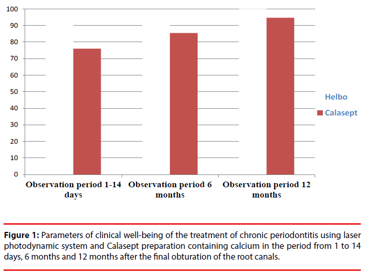
Treatment results were evaluated in the period from 1 to 14 days, 6 months and 12 months after final root canal obturation. Parameters of clinical well-being of the treatment of chronic periodontitis using laser photodynamic system and Calasept preparation containing calcium are shown in Figure 1.
It should be noted that clinical well-being in the nearest terms was observed in 95.5% of patients of group 1 and in 76% of patients in group 2. Complications occurred in patients of group 1 in 4.5% of cases. In group 2, complications were found in 24% of cases, which is almost five times higher than in group 1. When comparing 2 methods of treatment of chronic periodontitis after 6 months, clinical well-being was found in patients of group 1 in 97.3% and in 85.5% of cases – of group 2. The percentage of complications in patients of group 1 was 2.7% and in the 2nd group – 4.5%. Thus, complications in patients of group 2 were observed almost 2 times more often than in the 1st group. When comparing two methods of treatment of chronic periodontitis after 12 months, clinical well-being was observed in patients of group 1 in 100% of patients, of group 2 – in 94.7% of cases. Complications in patients of group 2 amounted to 5.3%.
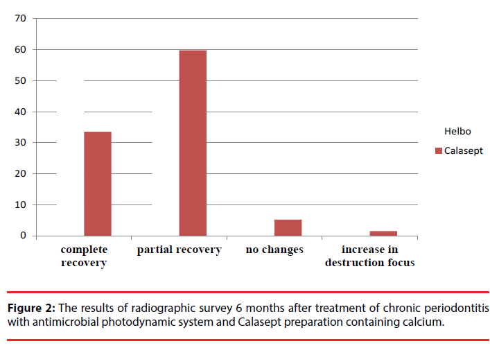
Radiological studies were conducted within 6 and 12 months to determine the degree of recovery of periapical exhaustion foci. Comparative evaluation of the results of radiographic parameters after 6 months in both groups is shown in Figure 2.
After the treatment for 6 months in both groups focus, recovery of periapical focus in bone tissue was observed in 50.3% of patients in group 1 and in 33.5% of patients of group 2. Partial restoration of bone tissue was found in 47.7% cases in patients of group 1 and in 59.8% of group 2. No changes in x-ray picture happened in patients of group 1 and in 2% of cases and in 5.2% of patients in group 2. It should be noted that in one patient in group 2, there was an increase in periapical focus, it was 1.5%. It was directed for surgery of resection of the root apex.
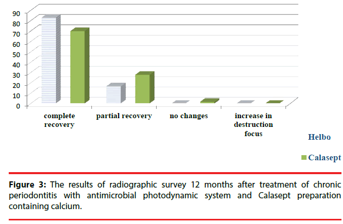
Comparative evaluation of the results of radiographic parameters after 12 months in both groups is shown in Figure 3. After the treatment of 6 months in both groups focus, recovery of periapical focus in bone tissue was observed in 83% of patients in group 1 and in 70% of patients of group 2. Partial recovery of bone tissue was found in 17% and 28.1% of cases, respectively. No changes in x-ray picture were observed in one patient from group 2 (1.9%). The patient is subject to dispensary follow up.
Conclusion
By analyzing the research results, we can conclude that the use of antimicrobial photodynamic therapy resulted in a reduction of clinical cases involving pain reaction after single stage of treatment of chronic periodontitis compared with data of patients treated with calcium hydroxide preparation.
Thus, the use of laser radiation in the course of preparation of the root canal for obturation when treating chronic periodontitis allows reducing the number of complications almost 1.5 times accelerates recovery of bone destruction foci. Furthermore, the analysis of observations of patients at different times showed that the introduction of laser disinfection of the root canals in the complex of endodontic procedures allows reducing the number of patient visits to the dentist, and allows conducting endodontic treatment per one visit.
References
- Maksimovsky Yu M. Endodontics and preservation of the tooth functions. News. Dentistry6(1), 3-6 (2001).
- Borovsky EV. Waiver of filling of the root canal by single paste –urgent task of endodontics. J. Clin. Dent 4, 18-20 (2000).
- Kodylev AG, Shumsky AV. Application of erbium-chrome laser in complex treatment of periodontitis. Endodontics. Today1,36-40 (2008).
- Dieva MB. Efficiency of endodontic treatment by method of depophoresis using “Medixid”. Cand. Med.ScMoscow. 22 (2005).
- Kunin AA, Erina SV, Pankova SN, et al.Modern aspects of endodontic treatment of the teeth. Clinical. Stomatology 1,18-19 (2003).
- Makeyeva IM, Zhokhova NS.Mistakes of endodontic treatment and ways to eliminate them. Central Research Institute of Stomatology – 40 years: history of development and prospects. 76, (2002).
- Mashchenko IS, Skotarenko AV. Treatment of chronic destructive forms of periodontitis with cyclophosphan. Dental. Market2, 62-67 (2005).
- Galanova TA.Comparative antibacterial activity of METAPEX and APEXDENT preparations in treatment of chronic periodontitis. Proceedings of the VII All-Russian Scientific Forum “Stomatology 2005”, Moscow. 62-63(2005).
- Pimenov AB. Segments of the root canals not available for tooling. Endodontics. Today3, 23-25(2003).
- Haapasalo HK, Siren EK, Waltimo TM, et al.Inactivation of local root canal medicaments by dentine, an in vitro study. Int. Endod. J33 (2), 126-131 (2000).
- Antanyan AA. Endodontic treatment per one visit: modern standards. Part 1. Endodontics. Today1, 78-84 (2008).
- Mitronin AV, Tsarev VN, Yasnikova E, et al.Features of contamination of the ecosystem of the root canals at the stages of endodontic treatment of acute periodontitis. Endodontics. Today1, 26-32 (2008).
- Schoop U, Kluger W, Moritz A,et al.Bactericidal effect of different laser systems in the deep layers of dentin. Lasers. Surg. Med35 (2), 111-116 (2004).
- Moritz A, Gutknecht N, Goharkhay K, et al. In vitro irradiation of infected root canals with a diode laser: results of microbiologic, infrared spectrometric, and stain penetration examinations. Quintessenc. Int28 (3), 205-209 (1997).
- Arazashvili LD.Treatment of chronic apical periodontitis with laser radiation. Urgent Questions of Endodontics. Proceedings of CNIISMoscow. 114-115 (1990).
- Friedman S. Orthogradic revision of the root canals: from concept to practice. Part one: prevention and treatment of complications after endodontic treatment. Dentali. Q1, 8-19 (2004).
- Kholenkov D. SiroLazer diode laser system: efficiency and compactness. Maestro. Dentistry3, 21-24 (2006).
- Moritz A, Gutkneht N, Goharkhay K,et al.In vitro irradiation of infected root canals with a diode lazer: Results of microbiologic, infrared spectrometric, and stain penetration examinations. Quint. Int28, 205-209 (1997).
- Prati C, Selighini M, Ferrieri P, et al.1994. Scanning electron microscopic evaluation of different endodontic procedures on dentin morphology of human teeth. J. Endod 20 (4), 174-179.


